Wednesday, September 4, 2013

Thermoregulation in horses in a cold time of year. Revised

by ©Natalija Aleksandrova
(Updated, presented at the 10th International EAHAE Conference, Poland, 2014)

Most horse owners are aware of the damage and crisis inherent with fever states. Few horse owners realize how well adapted horses are to deal with cold when certain aspects of their lifestyle are in place for them.

In order for a mammal to survive, internal body temperature is kept within a very narrow range. If the temperature exceeds these limits either above or below, the chemical reactions in the body function improperly, or they stop functioning at all. Fluctuations outside of the normal temperature range result in health problems or death of the animal.

Mature horses maintain their internal body temperature at a range of around 38℃. Foals, rapidly growing youngsters, pregnant and lactating mares have a higher than normal internal body temperature (Hines, 2004).

Heat in the horse's body is continuously generated as a by-product of metabolism, and a healthy animal has significant internal sources of heat from the metabolic processes (Bicego at al., 2007). To control internal heat loss during the cold time of year, the horse is provided by Nature with complicated and extremely efficient anatomical, physiological and behavioral thermoregulatory mechanisms.


On a genetic level, the domestic horse is the same as its wild counterpart of ancient and of modern times: it has the same abilities and needs to survive and thrive. In order for their amazing natural thermoregulatory mechanisms to be used in the most efficient way, or at all, the domestic horse requires nothing more from the human than only to provide living conditions which respect their natural needs — species appropriate living conditions. These are conditions which fulfill all the essential biological needs of the horse and allow it to exhibit its natural behaviors which have evolved over several thousands of years:
· herd life/social life (the horse is a herd animal; its brain has different capabilities which the brain of solitary animal does not have; only in a horse living in a herd, its cerebral cortex works properly, giving normal correct orders for functioning of other subordinated brain centers, only such horse is sound and psychologically balanced);
· freedom of movement 24 hours a day (movement is horse's metabolism; hoof health depends on movement);
· free access to forage 24 hours a day, grazing and/or hay (the horse stomach secretes stomach acid non-stop throughout day and night; it needs to be buffered constantly not to develop ulcers)
· free access to shelter, either built or naturally occurred, which first could serve as wind breaker;
· optimal hoof care, either natural or physiologically correct trimming.

We will see how the thermoregulatory mechanisms work in the horse, and how it can be interfered with and damaged through unnatural care and keeping practices when the animal becomes a subject for anthropomorphism. And very importantly, we will see how the horse doesn't always need to grow very long winter hair to feel comfortable during cold times. Long thick coat is just one of the thermoregulatory mechanisms of the horse, but NOT the only one.

Due to some thermoregulatory factors such as the skin and coat being very good insulators which prevent heat loss and the muscles producing heat through their movements, it is far easier for horses to warm up in cold weather than to cool down in hot weather or after intensive exercising. Cooling down is more difficult for the horse. Horses are adapted to handle cold.


Domestic horses taking bath on a spring day after snow just has melted, helping this way their winter coat to shed.

Skin

The horse's skin is responsible both for protecting the interior of the body from outside temperature changes, as well as for not allowing heat loss in cold weather. The skin is also responsible for dissipation of internal heat generated by muscle action in order to prevent the body from over-heating. The skins' thermoregulatory mechanisms consist of four major factors: skin itself, coat, arteries and sweat glands.

1. The skin itself works as an insulating layer through its relative thickness.

2. The coat.
The coat insulation depends on the depth and thickness of the hair layer, the wind speed and the temperature and humidity gradients within the coat (Ousey et al., 1992).

The horses coat changes twice a year through the mechanism called photoperiodism, adapting to different seasonal base temperatures. Sensors in the horse's skin react to the daytime light length changes. The horse is ready to grow their winter coat right after the summer solstice, when days start becoming shorter. The horse is ready to change their winter coat to a summer one right after the winter solstice, when days start becoming longer.

In addition to photoperiod, environmental temperature also affects hair growth. Colder climates produce thicker and longer coats than warmer climates do, when comparing horses who have the same body score and are fed the same amount of food.

Fetuses and newborn foals are provided with the mechanism which controls their coat growth as well. We can see that foals born in early spring are born with a longer coat than those born at the end of spring or in summer.


In addition to growing its coat, the horse can increase the insulation of the coat through the mechanism called piloerection — the raising, lowering or turning in different directions of each individual hair in the coat via hair erector muscles. In this way the horse increases or decreases the thickness of the insulation layer and efficiently varies the amount of airflow to the skin surface. Piloerection increases coat depth 10% to 30% in mature horses (Young & Coote, 1973).

<
Coat in an arabian breed horse on a very cold winter day (around –27˚C/–17˚F), Central Europe. The piloerection mechanism in use — the hair is raised to increase coat insulation.

The hair erector muscles must be exercised regularly in order to work properly, as with any other muscle in the body.

Hairs of the coat are covered with a greasy substance which creates a water-repelling effect that helps prevent moisture from reaching the horses skin on rainy or snowy days. Water runs down the outer hair while the deeper coat remains dry. Also body heat released via the skin helps to keep it dry inside.


Through regular coat brushing and shampooing, the greasy substance gets removed and the water-repelling effect becomes impaired.

It is not advisable either to clean off the layer of dirt that a horse gets from rolling in mud. Besides being protection against insects, the mud has a cooling effect in warmer weather.


Needless to say, that the popular practice of clipping the hair of a horse's coat completely eliminates the thermoregulatory factor of the coat.

3. Arteries in the skin.
Arteries through muscle actions, called vasoconstriction or vasodilation, can be narrowed or enlarged, regulating blood flow to the skin.

Constricting prevents internal heat loss by reducing the amount of warm blood brought to the cooler body surface. Dilation allows for a larger amount of hot blood from over-heated interiors to reach the body surface and to be cooled. The cooled blood lowers internal body temperature when it's returned back to the interior of the body.

4. Sweat glands.
When the outside temperature is too high for the air to cool the blood through the skin, the sweat glands secrete fluid. Evaporation of this fluid cools the skins surface as well as blood in the surface arteries. In this way, the internal temperature can still be lowered even when it’s hot outside.

The horse may also use sweat glands in extremely cold weather when internal body temperature is too high from exercising.

The horse stops secreting sweat as soon as the internal body temperature has reached its normal range. It then must dry quickly to avoid over-cooling. For this a sweaty horse turns its coat hairs in various directions. If given freedom, it usually seeks a windy spot to quickly, safely and effectively dry itself, contrary to human belief that a horse will catch a cold if stays wet in the wind.

Mentioning the sweat glands mechanism is important because sweat glands are also brought into function through muscle action.

When the body is hot, but not enough to secret the sweat, the horse may use rolling in snow.


Now let's look into other thermoregulatory mechanisms available to the horse.

Lungs

In addition to the skin, the coat, changes in blood circulation and sweating as means of controlling internal temperature, the horse has access to a cooling mechanism involving the lungs. Air exhaled from the lungs contains moisture. In warm conditions, if the above thermo-regulatory mechanisms are not adequate to cool the body, the horse can increase the breathing so that more air is taken in to the lungs and more (warm) water vapor is expelled – taking heat from the body. Most of us are familiar with dogs panting to cool themselves.

Increased breathing can be an effective way to cool the body but unfortunately many horses already breathe more air than is good for them. This is because factors such as stabling, isolation, unnatural eating habits, blanketing, clipping, shoeing etc., impose stress on the physiology which involves increased adrenaline production which is linked to an increase in the volume of air breathed. Over breathing has a direct and damaging effect on the physiology (including for example reduced oxygen availability), and at some point, the physiology may be unable to function adequately and then symptoms arise (http://www.equinebreathing.com).

Over breathing in a cold time may cause excessive loss of internal heat.

Body fat

The amount of fat in the body is another important factor of thermoregulation. In addition to being the body's energy reserve, fat is 3 times more insulating than other tissues due to its low thermal conductivity and poor blood supply (Guyton, 1991; Davenport, 1992). Thus it is important for a horse to have a good layer of fat before winter.

Wild horses and naturally kept domestic horses maintain the natural rhythm of weight change throughout the year with their weight growing up to 20% by Autumn.


Usually we can see that domestic horses with a thicker fat layer in their bodies grow a comparatively shorter winter coat than horses with less fat, when comparing the same breed and size animals. We often see an excessively long hair coat in ill or old animals who have trouble to keep weight due to pain, teeth problems, etc.; or in neglected underfed animals.

Also fat gets distributed more evenly over the body surface in cold conditions instead of being concentrated in some particular areas as it does in hot conditions.

In general, this is one of the cases where a horse seemingly 'doesn't grow enough coat' — it is a horse with a higher body score, who compensates for a longer coat with the body fat.
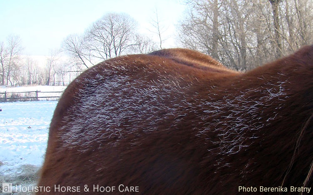

The horse with a thicker fat layer — we can see the excess heat escaped the body and is visible as the frost on the coat.

Size/shape of the body

Kept in the same conditions, smaller horse breeds have a longer/thicker coat compared to larger breeds. Also we typically see a thicker coat in foals. This is connected to a great effect of allometry on heat balance within animal species. (Allometry — the systematic change in body proportions with increasing body size.) Changes within species occur as animals grow and develop but exist also between breeds of species (Reiss, 1991; Langlois, 1994).

Generally, large body size is an advantage with respect to thermoregulation in the cold. Since, the ratio of heat-dissipating surface area to heat-producing/retaining body mass decreases with increasing body size (Phillips & Heath, 1995; Bligh, 1998). Therefore, large size horses have less relative surface area available for heat exchange, and thus importantly lose less heat in the cold than small size horses do. Small horses lose more body heat than large horses do.

In addition to large body size, a spherical body shape reduces the surface area to body mass ratio (Langlois, 1994). To compensate for the bigger surface/mass ratio northern-type horses, native breeds and ponies generally have evolved heavier rounder bodies with shorter limbs and extremities which are well protected by thick hair, mane and fetlock. Therefore they are more able to retain more body heat and cope with the cold.

Another possible reason your horse 'doesn't grow enough coat' — she is an 18 hand warmblood.

Digesting fiber

Increasing feed intake increases heat production in the horse's body. This is connected to the fact that the process of digesting long fibers produces heat as a by-product.

In cold weather we can observe an increase of food intake in horses. Such extra demand for feed is called climatic energy demand (MacCormak & Bruce, 1991). Horses have been observed to need up to 2.5% more energy for maintenance per 1 degree Celsius drop in outside temperature below their lower critical temperature (Young Coote, 1973; McBride et al., 1985; Cymbaluk et al., 1989a; Cymbaluk, 1990). (Lower critical temperature is individual for every horse/group of horses at different times of year and depends on many other thermoregulatory and environmental factors.)

Importantly, smaller-sized horses have greater low critical temperature values. Thus small-sized horses actually need proportionally more additional feed per kg of the body weight, than bigger-sized horses.

With this thermoregulatory factor, the need of the horse to have free access to food 24 hours per day throughout a year becomes especially important. In colder weather it gives it a chance of increasing heat production through continuously consuming and digesting long fiber. Especially when some of the other thermoregulatory mechanisms aren't yet adjusted in suddenly changing weather conditions such as a rapid drop of temperature.

And here it is important to note that all kinds of slow feeders or feeders preventing a horse from having mouthful of hay whenever it wishes, are not quite natural for the horse. This way of feeding cannot be really considered as fulfilling the horse's need for free access to food as it is in the wild.


Wild mustangs in Twin Peaks HMA, California, USA. Spring in the range grasslands — lush green grass is mixed with last year dry grasses.


Przewalski horse in Mongolian steppe, summer.

Dry winter grass still can be quite high in sugars and other nutrients, it is not the same as straw.


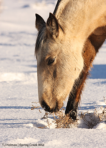
A domestic horse refreshing his menu with old dry grass in winter.

Reducing activity in cold

Feral horses have been reported to reduce locomotor activity in winter compared to summer (Duncan, 1980; Berger et al., 1999; Arnold et al., 2006). Reduced activity in winter is an annual pattern related to decreased outside temperature and thus to a reduction in internal heat production and spending energy (Arnold et al., 2006). This adaptation mechanism of reducing activity helps horses to cope with the energetic challenge of winter.

We can observe similar reduction of activity in winter in domestic horses kept in species appropriate living conditions. Even though the domestic horses usually aren't challenged with a necessity to search for food in winter, this slowing down in their activity obviously has the same purpose as in the wild horses — the reduction of energy wasting in the cold. Thus, it is a normal seasonal rhythm in the horse to exercise less in winter, therefore it is not advisable to forcefully exercise horses in winter.

Short-term activity in cold

Along with general reduction of activity in the cold, we also can observe short sessions of restlessness and locomotor activity during sudden acute cold periods and adverse weather. Short term beneficial movement is a useful bridge until other factors of their thermoregulatory system adjust to the new temperature conditions.


Reducing heat loss and gaining body heat via body radiation

Sometimes we can observe horses standing or lying down very close to each other. This way they reduce heat loss via radiation. By such positional closeness to each other they reduce the body surface area exposed to the external environment (Bligh, 1998). At the same time, animals who for some reason don't produce enough individual internal heat can use a paddock mate's body-heat radiation via positional closeness as an extra source of heat.


'Sun-bathing'

Also by changing body posture and orientation, horses can increase absorbed solar radiation as another additional source of heat.

Often we can observe that horses prefer to sunbath under the direct sun instead of eating on short sunny winter days, and as soon as the sun sets they are back to eating. This way they accumulate the sun's energy. This helps them to stay warm without using own body energy.


'Sun-bathing' on a winer day.


Body position/posture

On windy, rainy, snowstorm days, we can see horses standing with their tails to the wind and their heads low. This way they effectively keep their necks, heads, ears and eyes, underbelly and sheaths out of water and wind. Their tails serve to protect their rear ends — the shorter hairs on the dock fan out deflecting both snow and wind.


Also on such days, horses can be seen standing by the walls outside shelters, or using natural windbreaks such as trees or hills to protect themselves from the elements. And sometimes using each other's body as windbreaker.


The filly uses the body of the older mare as a wind breaker.

When allowed free choice, it's been observed that horses utilize enclosed spaces, such as shelters or forests, mostly to hide from summer heat and flies.

Snow on backs

Snow which we can sometimes see lying along horses backs during winter also plays the helpful role of providing an extra protective layer against internal heat loss. The snow melted underneath and frozen on top, creates an extra protective layer in a snow storm.


Shivering

Under extreme circumstances, heat in the horse body can be generated by shivering. During shivering, heat is rapidly produced by breaking down ATP* in the muscles (Langlois, 1994). Shivering is usually an acute response to sudden cold exposure. Or sometimes it occurs during extended periods of exposure to cold in rainy weather. In healthy animals, shivering is replaced by normal internal heat production as they adapt to new weather conditions.
(*Adenosine triphosphate — transports chemical energy within cells for metabolism)

Thermoregulation in the hooves

A constant temperature in the hooves is needed for proper metabolism to take place, which with the help of a few other factors, allows for normal horn production. The hard hoof capsule itself has insulating properties. Through hoof mechanism — the expansion and specific deformation of the hoof capsule and inner structures on impact — the impact energy created which deforms the hard hoof capsule and its inner structures, releases heat as a by-product thus keeping the hoof warm. For hoof mechanism to function properly, a horse needs unrestricted hooves, as well as unrestricted movement throughout the day and night. Affixed metal shoes impair the hooves ability to keep this constant temperature inside the hoof capsule by blocking the ability to expand and deform properly on impact. The nails also conduct cold deep inside the hoof capsule. We can often observe how snow builds and balls up on the soles of shoed horses, where as normally (on healthy uninhibited hooves) it would melt.

Effects of blanketing

Blanketing a horse causes a complete mess of the thermoregulation system. A horse cannot increase heat in selected areas of the body. So when trying to warm up parts of the body left exposed to the cold such as the head, neck, belly and legs, the horse in the process becomes over-heated in the parts covered by the blanket. The whole body cools or the whole body heats up. Sweating under a blanket is more of a problem metabolically to the horse than people realize.

What happens if a hot sweaty horse is placed in a stable?

Due to a lack of air circulating in enclosed spaces, cooling takes longer and the horse sweats for longer. The air surrounding the horse becomes saturated and therefore drying also takes longer than normal, because the humid air cannot absorb any more moisture. As a result, the horse remains undercooled, setting the stage for internal disorders: colic, diseases and infections by negatively affecting metabolism's safe temperature margins.

What happens if a horse is continually kept in stables and/or blanketed

Kept in stables or/and blanketed, horses lack stimuli (such as temperature fluctuations [flak…]) triggering the activity of thermoregulatory mechanisms. They don't need to exercise hair erector muscles, nor to dilate or constrict arteries, nor to activate the sweat glands, nor to prepare or deplete healthy fat reserves. All muscles atrophy without exercising after a period of time.

If an animal in this state is suddenly exposed to the cold, they will not be able to activate necessary thermoregulatory mechanisms. As a result the internal body temperature could drop too low which would lead to disruptions in metabolic processes. This can affect, for example, the production and migration rate of white blood cells and antibodies, with partial disabling of them. The result is a stressed animal with a disease or infection hosting internal environment. "The germ is nothing, the terrain is all". Consequentially germs or viruses in the body are allowed the perfect opportunity to over breed.

Stress effect

Besides the fact that the natural thermoregulatory mechanisms can only be fully utilized when a horse is kept in their species-appropriate living conditions, there is an anxiety and stress factor that horses inevitably experience when cut off from their basic needs and are kept in ways unnatural for this species (stabling, separating from equine companions, forced exercising, lack of continuous fiber intake, etc.). The stress also makes them less capable of coping with colds. We know it from human medicine that stress weakens the immune system.

Strengthen your horse immune system giving her the life she was designed for by Nature!


This writing was inspired by researches of and dedicated to one of my most important teachers — Doctor Hiltrud Strasser.

_____________
References

Arnold, W., Ruf, T., & Kuntz, R. (2006). Seasonal adjustment of energy budget in a large wild mammal, the Przewalski horse (Equus ferus przewalskii). The Journal of Experimental Biology, 209, 4566–4573.
Autio, E. 2008. Loose Housing of Horses in a Cold Climate. Doctoral dissertation. University of Kuopio, Kuopio, Finland.
Bicego, K.C., Barros, R.C.H., & Branco, L.G.S. (2007). Physiology of temperature regulation: Comparative aspects. Comparative Biochemistry and Physiology, Part A, 147, 616–639.
Berger, A., Scheibe, K-M., Eichhorn, K., Scheibe, A., & Streich, J. (1999). Diurnal and ultradian rhythms of behaviour in a mare group of Przewalski horse (Equus ferus przewalskii), measured through one year under semi-reserve conditions. Applied Animal Behaviour Science, 64, 1–7. Press.
Bligh, J. (1998). Mammalian homeothermy: an integrative thesis. Journal of Thermal Biology, 23, 143–258.
Cymbaluk, N.F. (1990). Cold housing effects on growth and nutrient demand of young horses. Journal of Animal Science, 68, 3152–3162.
Cymbaluk, N.F., & Christison, G.I. (1989a). Effects of diet and climate on growing horses. Journal of Animal Science, 67, 48–59.
Davenport, J. (1992). Animal life at low temperature. London, UK: Chapman & Hall. Duncan, P. (1980). Time-budget of Camargue horses II. Time-budgets of adult horses and weaned subadults. Behaviour, 72, 26–49. ogy, 163 (7), 602–607.
Equine Breathing. How well does your horse breathe? (http://holistichorseandhoofcare.blogspot.com/2014/10/how-well-does-your-horse-breathe.html)
Guyton, A.C. (1991). Textbook of medical physiology. 8th ed. Philadelphia, USA: W.B. Saunders Company.
Hines, M.T. (2004). Changes in body temperature. In S.M. Reed and W.M. Bayly (Eds.). Equine internal medicine (pp. 148–155). St. Louis, USA: Elsevier.
Langlois, B. (1994). Inter-breed variation in the horse with regard to cold adaptation: a review. Livestock Production Science, 40, 1–7.
MacCormack, J.A.D., & Bruce, J.M. (1991). The horse in winter — shelter and feeding. Farm Building Progress, 105, 10–13.
McBride, G.E., Christopherson, R.J., & Sauer, W. (1985). Metabolic rate and plasma thyroid hormone concentrations of mature horses in response to changes in ambient temperature. Canadian Journal of Animal Science, 65, 375–382. 187–194.
Ousey, J.C., McArthur, A.J., Murgatroyd, P.R., Stewart, J.H., & Rossdale, P.D. (1992). Thermoregulation and total body insulation in the neonatal foal. Journal of Thermal Biology, 17 (1), 1–10.
Phillips, P.K., & Heath, J.E. (1995). Dependency of surface temperature regulation on body size in terrestrial mammals. Journal of Thermal Biology, 20 (3), 281–289.
Reiss, M.J. (1991). The allometry of growth and reproduction. Cambridge, UK: Cambridge University Press.
Strasser, H. 2000. A Lifetime of Soundness. 3d ed. Published by S. Kells in Canada.
Young, B.A., & Coote, J. (1973). Some effects of cold on horses. Horse report at Feeders’ Day. Alberta, Canada: University of Alberta, Department of Animal Science.
__________________

English Edit Courtesy:
Jamie Joling, 2014
Tamlyn Labuschagne Ennor, 2012
______________________

For publishing permission, please send alHHHC a message on facebook: https://www.facebook.com/alHHHC

Thursday, August 8, 2013

Does my vet know?

by Maksida Vogt, Natalija Aleksandrova

In these times as we are becoming increasingly educated on different aspects of the health of our horses, and animals in general, quite a few situations arise which cause us to think deeper about the knowledge and treatment options that veterinarians apply to our animals. This article is not meant to be an affront to the knowledgeable and responsible up-to-date veterinarians. The sad fact however is that we routinely encounter a pattern that an international majority of veterinarians display. The pattern is a lack of knowledge about horses biologically correct keeping, about illnesses connected to biologically wrong keeping, about the natural normal behavior of these animals inside of and outside of their biological niche, as well as treatment of simple infections and abscesses related to their biological niche requirements being disregarded.

This problem is not connected to any particular country. In fact, we watch it happening in many places, as our organization receives feedback from horse owners of different countries, and our professionals encounter it on an ongoing basis everywhere as well. It seems this is a general problem. With this article we would like to encourage horse owners to never be afraid of questioning the proposed work of veterinarians and the health of their horses.

The scientific thesis by Dr. Cook, which conclusively proves the harm of the bit in the horse’s sensitive mouth, has already been in print and available for YEARS, it is however yet to be included in veterinary school syllabus, and is largely unknown by vet professionals. The bit is a foreign body laying on the mucosal tissue and the nerve endings of the jawbone. In the mouth of a horse the presence of this foreign body, alone, triggers the chewing reflex, and the horse starts producing saliva. Influencing this foreign body in different ways via the rider’s hand causes neurological discomfort and pain. We have an amount of photographs documenting the damages caused by bits, starting from bruising up to and including severe tissue trauma of tongue, soft palate and damaged mandibula, problems with teeth. These documented proofs were published in scientific magazines and to date remain unopposed, yet veterinarians still do not use this knowledge in their practice and do not educate horse owners regarding this problem. This leaves room for speculation on the quality of their knowledge and also on possible reasons that might influence their lack of action.

Especially regarding hooves – most veterinarians lack up-to-date knowledge on diagnosis and correct treatment options. Here we would like to present several cases of incompetency in the diagnosis and treatment of different hoof problems, which we collected from horse owners and our practice.

Case 1. A wrong treatment of hoof problems by a team of veterinarians and a farrier

A mare 19 years of age, was rescued and arrived with a so called „orthopedic“ shoe on one of her front hooves. The treatment was for ‘hollow foot’ (‘white line disease’ with the wall separation), a problem which she had had for years. The mare was a competition show jumper with this foot — an unbelievable fact, which gives us another serious reason to question the existence of equestrian „sport“ and people who are in it and who support it. The owners/rescuers were told that this problem was ‘nothing special’, but in reality the hoof could not keep even a shoe on anymore. The mare needed to be transported to a clinic at Telgte. At this clinic a farrier pushed a nail into the hole in the hoof wall to see how deep it went, a softer object would have sufficed. Then he performed a hoof wall resection, removing a large section of the dorsal part of the wall.


The mare was sent home with a bandaged hoof and with a plastic pad underneath serving as „protection“. It was advised that the mare was to be kept confined in a box rest situation. The mare spent one year in the box. Her bandage was renewed every few days. Everything was done exactly as prescribed by the veterinarian team. The owners followed their advice in the belief that the team knew what they were doing and that they were in fact helping this mare. The owners made considerable efforts to give the mare more space in which to live in her Solitary confinement by offering her the space of two boxes and a small paddock. After the year, when nothing had improved and the hoof wall still remained separated, the owners decided to reject further advice from this team of vets and farrier and to search for another solution. They decided to let this horse out of the box so that she could move freely together with the herd. Their decision proved to be a sound one as they observed over time that this free movement situation alone contributed to the improvement of her hooves and better growth of the hoof wall.

Case 1. Analysis

Despite the wide spread believes, bacteria and fungi do not create any threat to the white line of a healthy hoof, since in the healthy situation the rate of horn production always exceeds the rate of its decompression. Bacteria and fungi are always present in any hoof, they are necessary to the hoof. When a healthy balance exists, fungai and bacteria are an integral part of the natural self-trimming mechanism of the hoof. (more: “Hoof thrush” >>).

The true cause of white line problems actually lies in incorrect hoof form and/or in shoeing and/or in improper horse keeping conditions, which restrict horse’s movement, cause hoof dehydration and horn damage via exposure to ammonia. The incorrect hoof form and/or shoeing and the lack of natural movement lead to reduced blood circulation as well as mechanical damage of the corium, which is the tissue that produces white line horn. These causes also lead to damage of the white line itself. Through the damage and poorer quality and quantity of the horn produced, the speed in which the horn is decomposed begins to exceed the speed at which horn is produced, and the white line is thus destroyed far up into the hoof capsule. The space now existing where the white line has been destroyed allows foreign bodies and infection to enter the interior of the hoof. Also the normal populations of bacteria and fungi, explode ‘out of control’ and they move deeper inside the space, ‘eating up’ the horn. If the hoof continues experiencing reduced blood circulation, and damaging mechanical forces continue affecting it, the laminar corium and the laminar horn get eventually affected, and the ‘hollow foot’ effect appears — the wall starts separating from the sole and the coffin bone.

In this situation, shoeing or so called ‘orthopedic’ shoeing can only mask the problem for some time as it remains the obstacle to healing, while the damage will continue progressing, often unnoticed by horse owners and professionals. Because, through restricting the hoof mechanism*, shoeing blocks the blood circulation in the hoof and creates damaging mechanical forces.

*(hoof mechanism >>)

The wall resection also doesn’t address the true cause, as its purpose is only to remove a part of a hoof ‘affected by bacteria and infection’. Quite contrary, the resection can create a situation where infection now severely affects the inner tissues and structure, as the hoof is cut open. Bandaging material and the placement of an artificial wall can create more damaging pressure on the corium tissues.

And the worst advice that can be prescribed in a situation such as this — when a hoof problem is caused by a lack of blood circulation, amongst the other — is putting a horse on the stable rest. It is a basic natural fact — healing can only happen through increased metabolism, i.e. through increased blood circulation. As “fresh” blood supplies a damaged area with nutrients — ‘building blocks’ needed to renew damaged tissue. The more intensive the circulation, the faster healing can happen. To put a horse on the stable rest means to reduce the circulation in her hooves, as the normal circulation in the hooves is only possible through the constant movement that the horse’s body and hooves are created for. Thus, restricting the circulation, we restrict the healing process.

Additionally, separating a horse from a herd, delays the healing process through resulting mental suffering and psychological imbalance. The horse is a herd animal and its brain has different capabilities than the brain of a solitary animal. The horse’s cerebral cortex is built to function normally giving normal correct orders to subordinated brain centers only when the horse lives in a herd.

First, what any veterinary practitioner should recommend in a case of white line problems is physiologically correct hoof care in proper keeping conditions providing unrestricted movement, which would promote the hoof mechanism and, through it, all the healthy hoof functions which are designed to promote healing.


Case 2. Wrong diagnosing and treatment of hoof problems by a veterinarian

A veterinarian was called to a mare who had been found lame in her right front leg the previous night upon returning from her paddock. The mare’s hooves were quite overgrown, and she was scheduled for a trimming session in a few days time. The mare had had her shoes taken off less than 2 months before and was sensitive on hard ground. The vet checked her lame foot and told the owner that the lameness was caused by laminitis in her right front because she had eaten too much grass. The vet took an x-ray of the one hoof, and from this evidence his next verdict was: 'O.k., no coffin bone rotation visible. It is not laminitis.' Though he still believed that the problem making the mare lame was in the hoof. The mare received painkillers and anti-inflammatory drugs and was put on a box rest. Two days later the mare walked normally, and was taken by her owner to the paddock and her herd, as she was becoming quite crazy staying inside alone.

Two days later there was still no visible lameness so the mare was trimmed. Day after trimming she was lame in her right front again. The vet came and concluded 'Laminitis'. He prescribed full rest, pain-killers and antibiotics. The mare stood all the time without putting weight on her right front foot. Loading her left front foot was not a problem. When walking she seemingly didn't have a problem in weight-bearing on her left front foot, but was limping on her right front foot. The owner couldn't agree that it was laminitis causing the lameness, and decided not to give all the prescribed medicine before she could feel sure of the diagnosis. Hoof care professionals in this stable also didn't agree it was due to laminitis, they believed it was rather more likely to be an abscess.

After a few days of the full rest, the owner took the mare outside for a short time. The mare was happy to move freely and trotted, though still lame. A day later the owner couldn't bear to see her horse suffering from separation anxiety from her herd and so she let her lame mare out into the paddock for night. The next day the abscess burst out in the area of the coronet. In a few days the lameness disappeared fully. The owner called the vet to inform it was the abscess. But the vet didn't agree the physical evidence. He still insisted it was laminitis in the right front. Later the owner got a bill from the vet, it stated on the bill 'Signs of the rotation of the coffin bone in the right front hoof visible on the x-ray. Diagnosis: laminitis.' (We remember that the owner was said 'no coffin bone rotation' in the beginning, don't we?)

Case 2. Analysis

Laminitis usually affects both of the front hooves or all four hooves in horses. The acute form of laminitis is characterized by pain a horse shows in all affected hooves. Laminitis can happen without pathological changes inside hoof capsule such as coffin bone rotation. In severe cases, the bone rotation can appear, but not before 48 hours after the signs of pain appeared.

The x-ray made the day after the signs of acute laminitis had appeared cannot be used as a confirmation of this diagnosis: it is too early for the changes to become visible. The changes visible on such x-rays can be the result of previous problems. X-rays may never show any changes, since the acute laminitis can happen without them.

Next, what might make us suspicious about the diagnosis 'laminitis' in this case is that it was diagnosed only in one hoof. It is extremely rare that a horse develops laminitis in only one hoof. So far the only conditions recorded under which such type of laminitis could occur: being a sequelae of excessive weight placed on a limb due to an injury to the opposite limb, when the horse is not allowed to move after being injured ('static laminitis', Rooney, J.).

Another statement, which contradicts the diagnosis 'laminitis in one hoof', is 'the mare got it because of eating too much grass'. All four hooves are ONE metabolic organ of the horse's body. The carbohydrate overload, which causes an effect in the body similar to intoxication, cannot affect only 1/4 of one metabolic organ in our case.

Now we look into the hoof shape of both of the mare's front hooves.


(Click to enlarge.)

We notice the same considerable degree of dorsal wall flaring in both front hooves, the flaring suggests to us a seriously compromised laminar connection similar for BOTH hooves. Having the same degree of damage of the lamina of both front feet, it is very unlikely laminitis could be triggered only in one hoof.

Knowing only these few facts could make us search for another explanation of the lameness in one hoof.

The horse was unshod not so long before the described events. De-shoeing can restore the hoof mechanism in the hooves to some considerable extent. With any restored hoof mechanism, the blood circulation is consequently also improved inside the hooves. With better blood supply, it becomes possible for the body to start repairing the damage, including cleaning the hooves from necrotic tissue, that was caused to the inner hoof tissues by the period of long time shoeing. Abscessing is a method the body uses to remove fragments of dead tissue, which are too big to be removed within the blood stream.

In this case lameness returned in all its strength directly after trimming. Removing the overgrown horn via trimming improved the hoof mechanism even more. Improved hoof mechanism means improved blood supply and thus improved healing capacity. The abscess, previously suspended in its function due to lack of blood supply was supplied new strength for resolving its work after the blood supply of the affected area was improved. Abscesses are supplied from the blood with all the tools they need as their work materials such as white blood cells. The better the blood supply, the more intensive a healing abscessing process can be.

This is why abscessing is not rare after de-shoeing and can appear right after just a planned trim.

Now we look again into hoof photos.


(Click to enlarge.)

If we look closer, we can notice 'seedy toe' in the right front hoof — the 'hole' in the toe. The constant pressure of the coffin bone tip into the sole due to an incorrect hoof shape (too high heels), combined with the pressure from the shoe clip lead to damage of blood vessels in this area and later to bone tissue destruction. Dead tissue from the bone and the corium was accumulating for a long time in this area. The damage in the horn created an entrance for infections from the outside. This creates the perfect situation for an occurrence of abscesses.

And if we look at this photo:


We can see the place, where the abscess eventually broke out at the coronet. It happened directly over the damaged spot in the toe. This is very typical behavior of abscesses — to find the shortest trajectory for exit through a more elastic horn, such as the laminar horn. Abscesses tend to come out at junctions of softer and harder horn.

(More on hoof abscesses >>)

Case 3. Unacceptable quality of x-ray shots made by a veterinary specialist and further mistakes in diagnosing hoof problems using the x-rays

A vet was called to an 18 year old gelding to make x-rays of his front hooves. The gelding had been shod for many years and de-shod a month previous to the visit. The x-rays of the front hooves were required by a hoof care professional for judging hoof conditions after de-shoeing. At the time of making the x-rays, the hooves looked like this:


These are the x-rays that were made for this case:


From the note the vet sent to the owner along with the shots, we learn amongst other things that '...the spacing between the hoof wall and the coffin bone seems to be bigger in the horse's right front hoof than in his left hoof, and this may indicate resorption (a loss) of the bone.'

Case 3. Analysis

The first and the very important problem, which the increased space between the bone and the wall indicates, is chronic laminitic changes inside the hoof capsule: damaged and weakened laminar corium, rotation with separation and sinking of the coffin bone. Even so that laminitis usually is presented in both front hooves, the degree of change can be different in each hoof due to differences in the shape of each hoof.

For the interpretation of bone loss alone to cause a visibly larger distance between the bone and the wall, it would have to be really dramatic bone re-modeling, which we don’t see in the bones on these x-rays. If the statement was to be correct then the kind of bone re-modeling the vet was talking about usually creates a specific shape in the dorsal surface of the coffin bone, making the extensor process look bigger, more pronounced than normal because, due to specifics of the hoof anatomy and physiology, it is not affected by the damaging pressure, which causes the bone loss in the dorsal surface of the coffin bone. We don’t see this specific shape in either bone on these x-rays.

The diagnosis states that only one bone is affected and that supposes a different degree of the remodeling to be shown in each bone, we don’t see such difference on these x-rays.

First what we actually notice about these x-rays is that their quality is unacceptable. They are unacceptable because no hoof soles are visible on them and the lowest part of the x-rays is overexposed. Thus we cannot judge by these x-rays such important things as thickness of the soles and position of the coffin bone in relation to the ground. Hoof x-rays should always be made with a visible ground level on the shot. There’s also no clearly marked junction between the wall and the coronet on the x-rays, this prevents one from correctly judging the degree at which the bones have sunk down.

There’s no significant difference in size of spacing between the wall and the bone between the two hooves visible on the x-rays. What we do notice, actually, is a difference in the coffin bone positions inside the hoof capsules due to the difference in the hoof shapes (we can see this difference on the photos) — one hoof is more upright (has higher heels) and the other is flatter. And we can see the degree of the bone rotation in each hoof.

When a not ground parallel coffin bone in an x-ray is visible, this is a certain sign of overload of the lamellae suspension and therefore in the most of all cases the reason for the laminitis (Glenn Ramsey, PhD candidate, New Zealand's University of Auckland, "The effect of hoof angle variations on dorsal lamellar load in the equine hoof," Equine Veterinary Journal, September 2011).

Obtaining similar x-ray results, a veterinary professional has to inform the owner of the danger of further progress of the pathological changes in such hooves and on a possibility of the horse developing acute laminitis if it doesn't receive correct hoof care, which would help to bring the coffin bone to a physiologically correct position inside the hoof capsules, and, through this, to relieve the laminar corium thus preventing further damage to it.

* * *
We do encourage the horse owners to ask all the questions they might have on this article and also on any treatment of their horses by veterinarians. We recommend following steps:

— Do not be afraid to question anyone for the sake of your horse.
— If you have an expert, then this expert must be able to explain every detail you ask for and also all the coherence regarding present problem in the horse's body. Bear in mind that conventional veterinary care is not holistic care and your vet might not know about holistic care.
— Refuse the treatment suggested by a vet who recommends that you shut your horse in the box. Horses need free movement to be able to heal.
— Refuse the treatment suggested by a vet who recommends chemical painkillers, corticosteroids or antibiotics for your horse without warning on their harmful side effects and without looking into alternative therapy possibilities (herbal, homeopathic, etc.). Besides causing metabolical chaos in the horse's body that makes the healing process difficult, chemical drugs are well known for their severe side effects in the horse’s body. Just a few of the side-effects: non-steroidal anti-inflammatory pain killers are known as a cause of stomach and gut ulcers; antibiotics are known to suppress immunity; corticosteroids are listed as a common cause of laminitis in horses.
— Refuse the treatment by a vet who does not consider any psychological side of the horse behavior (horse is a herd animal, separation from the herd causes enormous stress and makes the healing difficult).
— Refuse treatment that only makes your horse “functioning” in order that you can use it again quickly.
— Search for the horse experts who will work at the body’s pace with the real healing processes the body has, no horse deserves less than this.

The cases presentation and analyse by Natalija Aleksandrova
English editing courtesy Tamlyn Labuschagne Ennor

Wednesday, July 10, 2013

Restricted grazing — does it bring the desired effect in horses?

By Natalija Aleksandrova

Continue the topic on importance of grass grazing for the horse, started here http://al-holistichorseandhoofcare.blogspot.com/2013/05/by-natalija-aleksandrova-through-course.html, we will look into a new research of Prof Paul D. Siciliano and his team published recently. The research brings more detailed proof to the fact that restricting a grazing time for a horse to fight her obesity and other metabolic problems, only creates more stress to the metabolism and overall health. When horse's grazing time is limited, she is able to consume more than 3 times more dry matter than normally. Such intensive load of nutrients in a short time stresses the digestive system and the whole metabolism.

Besides the physical stress to the body, it also brings mental stress to the horse. Such horse can never be in peace in a pasture, expecting her time there runs out each moment, and also, if the grazing area is limited to too small, expecting it will be empty of nutrients soon. Also, if the grazing area is limited, it brings additional mental stress to the horses as higher in hierarchy horses continuously chases away lower ones in search for more nutritional grazing spots.

The research

While researchers reported long ago weight lost helps improve horse’s overall health, until now no one hasn’t known exactly what impact restricted grazing has on the equine gastrointestinal health or nutrients intake. A group of North Carolina State University researches, led by Prof Paul D. Siciliano, set a goal to investigate in more detail if restricted pasture access affected horse’s intake rate, energy intake, and hindgut fermentation.

The team separated eight mature idle geldings into four groups and allowed each group pasture access for either 3, 6, 9, or 24 hours for seven days. After seven days, the team reassigned the groups to a different turnout treatment on an ungrazed pasture. By the end of the four-period study, each horse had been subjected to each pasture treatment. When not on pasture, the horses stayed in drylot pens with access to water and salt; horses in the three- and six-hour treatment groups had free-choice access to a low-quality grass hay. Throughout the study, the team recorded the amount of hay consumed and collected fecal samples from each horse on each Day 7.

The team also measured or estimated pasture plant composition, herbage mass (used to quantify pasture available to an animal), grazing height, and forage preference for each period. They noted no difference in digestible energy concentrations (DE) or initial herbage mass for each pasture. However, less pasture was available during periods 2 and 3 compared to 1 and 4.

Key study findings included:
— Horses' total daily dry matter (DM) intake (pasture plus hay, if hay was provided) was not affected by length of turnout time. In other words, horses consumed the same amount regardless of the amount of time they were allowed to graze.
— The team found the highest total daily DM intake — which equaled 1.4% of body weight (BW) — in horses on pasture for 24 hours, which is less than the 2-3% BW previous research suggested. Siciliano and colleagues suggested the lower DM intake in their study could be due to high temperatures seen during portions of the research: High temperatures have been shown to decrease forage intake in horses up to 15-20%.
Pasture DM intake rate increased with restricted grazing. Horses grazing for only 3 hours had a higher intake rate compared to horses grazing for 9 and 24 hours, and horses grazing for 6 hours had a higher consumption rate than horses on pasture for 24 hours.

Amongst their other findings, the researchers also learned that horses' fecal pH — which is both influenced by diet and used as an indicator of hindgut pH — decreased as time on pasture decreased (lower pH means higher acidity); an acidic environment in the hindgut (termed hindgut acidosis) can lead to colic and other health concerns. This showed the team that length of time on pasture affected hindgut microbial fermentation. The authors believed the increase in rate of pasture intake could have played a role in the lower fecal pH, especially when the horses consumed high-quality pasture. However, they stressed, all fecal pH values were within the range considered to be normal.

Average DE intake was greatest when horses were on pasture for 24 hours, but total DE intake did not differ between treatments. The horses consumed 40%, 66%, 67%, and 94% of their total DE requirements with 3, 6, 9, and 24 hours of pasture access, respectively, which indicates that horses increase their rate of consumption with decreased time on pasture.

The team concluded that their findings support the belief that reduced time on pasture increases consumption rate and decreases fecal pH in horses.

“Simply reducing the time a horse spends at pasture may not always be an effective means of decreasing caloric intake,” said Siciliano. The team also noted that more work is needed to develop methods to accurately predict pasture intake of horses grazing for periods less than 24 hours.

The full study, "Effect of Restricted Pasture Access on Pasture Dry Matter Intake Rate, Dietary Energy Intake, and Fecal pH in Horses," is published in June in the Journal of Equine Veterinary Science.

***

If your pasture is specious enough to provide your horses with enough grazing throughout grazing seasons day and night, horses’ time in the pasture shouldn’t be limited artificially.

Also whenever it is possible, instead of closing your grazing area for recovering of grass there, when this area is limited, rather enough good quality hay should be provided for the horses additionally to grazing. This way the pasture will have a chance to recover naturally, at least to a certain extent, as the horses will choose naturally to eat more the higher quality hay, when the quality of the pasture grazing drops considerably due to overgrazing.

Horses, which are never limited in their grazing time have their life full and happy, finding a lot of other amusement in a pasture besides grazing.

For example, have a rest, including deep sleep:


Or play:


Communicate to each other:


Communicate to people:


Take care of the skin and coat:


Find a herbal help needed:


They move actively:


And, yes, they consume nutrients needed for life:


All photos except no. 12 by Berenika Bratny

Wednesday, June 5, 2013

The Importance of Species Appropriate Feeding and Eating Behaviour of Horses

by L.D.

Unlike the human’s digestive system, the horse's digestive system is designed by Nature to receive food in small portions almost constantly, day and night. By locking horses in stalls and making their feeding schedule similar to that of a humans — very few meals a day with long breaks in between — humans ruin the horse's organism, making the animal suffer from pain caused by the health disorders it develops as a consequence of the unnatural feeding schedule, such as gastric ulcers, colic, etc. In order for your horse to stay healthy and happy throughout its life, it should be provided species appropriate keeping conditions, which means conditions that are as close as possible to the natural way of life of the horse. This means all horses should be provided 24 hour access to grazing or/and hay, should be provided the possibility of unrestricted free movement 24 hours a day (the 'open stable' system) and be able to live together with equine companions. Now let’s take a scientific look into the importance of species appropriate feeding/eating behavior in horses.

__________________

This article focuses on the importance of species appropriate feeding/eating behaviour of equines. However, as also mentioned in the five freedoms (Farm Animal Welfare Council, 2009), which are building the base of the animal rights and protection, other factors are crucial in order to guarantee the well being of the horse:

1. Freedom from Hunger and Thirst — by ready access to fresh water and a diet to maintain full health and vigour.
2. Freedom from Discomfort — by providing an appropriate environment including shelter and a comfortable resting area.
3. Freedom from Pain, Injury or Disease — by prevention or rapid diagnosis and treatment.
4. Freedom to Express Normal Behaviour — by providing sufficient space, proper facilities and company of the animal's own kind.
5. Freedom from Fear and Distress — by ensuring conditions and treatment which avoid mental suffering.

Not only species appropriate feeding, but also being free to express species appropriate behaviour, such as herd-life, movement, mutual grooming and more, are of utmost importance and need to be fulfilled in order to guarantee the health and well being of the horses (and any other animal).

Scientifically speaking, equines are herbivore hind-gut fermentors. Having evolved to almost constantly eat little portions of long, rough fiber being low in energy. Observations and researches showed that horses being free to choose their feed stuff spend approximately 60% (~14 hours) of the day browsing and grazing (Gudmundsson and Dyrmundsson, 1994; McGreevy, 2004). Horses in a scarcer environment spend approximately 16–17 hours per day browsing and grazing, which may even exceed 19 hours in harsh seasons (McGreevy, 2004). However, the time spent eating strongly depends on several factors such as weather conditions, availability of feed stuff, gender, state of maturity and more. (Gudmundsson and Dyrmundsson, 1994).

Given the possibility to free range and express species appropriate eating behaviour the horse will consume approximatly 2–2,5% Dry Matter of their body weight within 24 hours, in the form of roughage. (National Research Council (U.S.), 1987; Kiley-Worthington, 1987; Dowler, et al., 2009; Wright, 1999).

Being able to free range and spend most of the day grazing results in approximately 57000 chewing movements (Cuddeford, 1999) on high fibre roughage (and about 40000 on roughage with an average fibre content, which in this research is the more likely number to work with) (Frape, 2004). Figures as such are often critically questioned as they are mainly derived from researches and observations from wild, feral or semi feral equines, however the genetic make up of ingestive related behaviour/instincts and physiology of the digestive tract have remained relatively unchanged over the last 6000 years of domestication and breeding (Koene and Gremmen, 2002; Dierendonck, 2006), which is then directly linked to the high occurrence of health and behaviour problems due to inappropriate nutrition.

The same eating behaviour performed by free ranging horses can be seen in domestic horses when given the possibility to execute this normal and instinctive behaviour (Kiley-Worthington, 1987). However, most domestic horses, nowadays, are only able to express limited normal eating behaviour due to human management. For example concentrates in the form of pellets, muesli or oats are fed in restricted meals with or without limited amounts of pre-cut hay or haylage. This change of feedstuff also manifests a change of eating behaviour in terms of eating time, chewing movements and, importantly, saliva production.

A horse being free to graze adequate mature pasture, 24/7, will consume an approximate Dry Matter intake of grass of 2 Kg per 100 Kg body weight per day (600 Kg horse). When being limited in grazing the horse will consume approximately 0,8 Kg DM of grass in one hour. The horse given limited hay at specific feed times will spend approximately 40 minutes per 1 Kg of hay, resulting in about 1,2 Kg Dry Matter intake per hour (Ellis, 2004). When given hay ad libitum it is most likely that the horse will spend longer eating the 1 ncentrates differs considerably; as the horse will only spend approximately 10 minutes eating 1 kg of concentrates (Ellis, 2004). This means that the horse has a high energy intake in a very short time. Which, firstly, the digestive system was not evolved for and secondly, the horse will spend much less time eating and chewing. This might result in a higher risk and occurrence of undesirable stereotypic behaviour and other health problems (Davidson and Harris, 2000) and might therefore influence the short and long term performance of the horse.

The horse's teeth have evolved for heavy wearing exposure and thus would need similar conditions in order to wear at normal rates (Baker and Easley, 1951). It has been observed that the change of the angle of the incisors, commonly in domestic horses is something which does not occur to such an extent in feral or wild horses. This means that abnormal wear takes place in many of the domestic horses. Such misalignments can have severe consequences on digestion and the whole equine body. As the tempro-mandibular joint becomes further misaligned it exerts a crucial role in altering the biomechanics of the whole body. Besides that, a change in the angle of the incisors means that the surface to surface contact of the molars is also put in an abnormal state forcing the horse to clinch its jaw in order to be able to chew feedstuff properly (LaFlure, 2001). Reasons for that abnormal wear are firstly, that the horse is fed with too little high fibre roughage. Secondly, it does not need to bite correctly anymore (reduced usage of the incisors), because the roughage is most likely precut. And, thirdly, the horse gets too much concentrated energy on which it needs to chew at least four time less than on hay (approximately 3000 chewing movements on 1 kg hay and 750 on 1 kg concentrates) (Frape, 1998).


As the production of salvia is linked to chewing (Alexander and Hickson, 1970) direct relations can and must be drawn. While eating a minimum of 14 hours a day and an amount of approximately 2–2.5% Dry Matter of the body weight, the horse produces approximately 4,5 litres of saliva per one Kilogram Dry Matter roughage, resulting in approximately 40–60 liters of saliva per day (Harris, 1999; McGreevy, 2004). Producing sufficient salvia is crucial for the buffering effect of stomach acidity. Saliva, with its high bicarbonate content, has an alkaline level and thus is able to balance the acid level in the stomach (Picavet, 2002). If insufficient saliva is produced at constant rates spread over the whole day, the buffering effects of the alkaline bicarbonate are diminished; the gastric acidity rises and risk of gastric ulceration increases considerably (Pagan, 1997). It has been observed that the occurrence of gastric ulceration happens most commonly in the upper region of the horse's stomach, as this part of the stomach does not contain a mucous membrane layer and neither produces alkaline bicarbonate as an acid neutralizer. The only protection that the upper region of the stomach has from gastric acid is the bicarbonate coming from sufficient saliva production and the natural buffering capacity of roughage (Pagan, 2008). Being exposed to a higher acidic level for more than three to four hours decreases tissue resistance dramatically, and the first signs of damage become visible after ten to twelve hours (Steward, 2003). Studies show that approximately 60% of performance horses and approximately 37% of leisure horses show gastric ulceration (Picavet, 2002). The highest occurrence of gastric ulceration is found in race horses. More than 90% of all race horses show gastric ulcerations to various degrees (Pagan, 2008). It has been indicated that horses would not voluntary starve themselves for more than four hours (Krazak, et al., 1991) as acid levels in the stomach increase by then; causing discomfort and initiating the desire to chew and graze, which effectively supplies buffering alkaline bicarbonate to the affected over acidic area.

Having evolved as a constant browser grazer also means that constant movement of the digestive tract is present and necessary to keep the micro environment healthy and active. The presence of beneficial bacteria prevents the spreading of other, potentially pathogenic bacteria (Pagan, 2008). If this constant peristaltic movement is not assured, the risk of colic (abdominal pain) increases considerably. In order to guarantee this peristaltic movement, the stabled horse being restricted in roughage intake will often be observed occupying itself with eating it's bedding, because it lacks fibre, saliva production as well as it needs to occupy it's time. Eating bedding is certainly beneficial for chewing, saliva production and time occupation and thus helpful in decreasing the risk of gastric ulceration, but straw bedding should not be the major source of roughage (it has almost no nutritional value and contains larger amounts of lignin, which is more difficult to digest.) However and importantly, excessive consumption of wheat straw bedding, has been implicated in the development of impaction colic (Thorne, et al., 2005; Cohen, et al., 1999).

Applying sub-optimal feeding regimes, not guaranteeing the natural requirements of almost constant eating, chewing, producing saliva and having a "full" digestive tract, does have severe consequences on physical and mental health. The development of gastric ulceration, colic, but also the development of abnormal behaviours, such as crib biting, (e.g. Kiley-Worthington, 1987; McGreevy, et al., 1995) might result and with that have a direct influence on the horses welfare, well-being and thus on the five freedoms (Waran, 2007).

English Edit Courtesy of Tamlyn Labuschagne Ennor 2012